TBI Blood Test GFAP: USMLE Guide to Biomarkers and CT
Clinician-scientists led by the University of Pittsburgh School of Medicine want to expand the use of blood biomarker tests after traumatic brain injury (TBI). The goal is to identify patients whose injury might otherwise be missed and to escalate their care. In a study in the Journal of Neurosurgery, the team reports clinical cutoffs for two brain-associated proteins, GFAP and UCH-L1, in patients with Glasgow Coma Scale (GCS) scores of 13 to 15. GFAP performed better than UCH-L1. The test is meant to add to clinical assessment, not replace it.
What happened
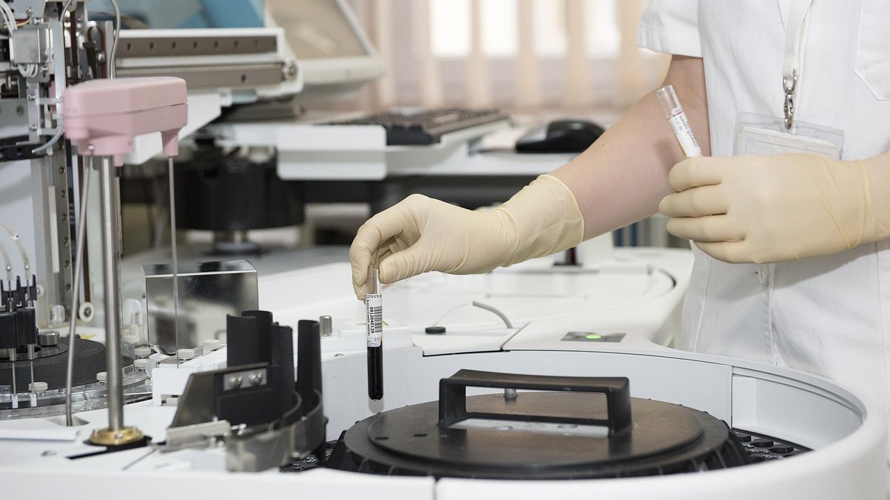
The study is part of TRACK-TBI, a multicenter, longitudinal project that collects data and biospecimens from patients with TBI who present to the emergency department (ED). The Pitt Neurotrauma Clinical Trials Center serves as the primary biorepository.
The researchers asked whether two blood proteins could do two things:
- Distinguish patients with mild TBI who had a positive head CT from those with a negative CT.
- Distinguish patients with a negative CT but a positive MRI from patients with both scans negative.
The two proteins were glial fibrillary acidic protein (GFAP) and ubiquitin C-terminal hydrolase-L1 (UCH-L1). Both rise in the bloodstream right after injury and then fall over time.
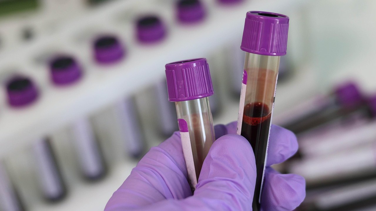
Within 24 hours of injury, blood GFAP could separate patients with acute brain injury from those with no imaging evidence of pathology. That group included patients whose MRI abnormalities were missed by CT. GFAP is released from glial cells and spikes sharply between 4 and 20 hours after injury, then declines slowly over the following days. UCH-L1, which comes from neurons, was less able to discriminate between the groups.
The authors framed the question in reverse of the usual approach. Existing tests help rule out the need for CT. Here, the team asked at what level clinicians could be fairly certain that a bleed is present, as well as at what level they could be certain it is absent.

Why it matters: CT misses some brain injuries
According to the source, about 2.8 million people in the U.S. experience a TBI each year. Earlier TRACK-TBI findings showed that more than 50% of participants with a mild brain injury had functional limitations at one year. The lead author suspects that one driver of poor outcomes is that patients are not properly identified in the first place.
Head CT is generally the first-line test. It is inexpensive, widely available, and fast. But roughly 25% of patients have intracranial pathology that CT misses. As the lead author put it, one in four patients sent home with a negative CT has an MRI abnormality suggesting they should have specialized follow-up. MRI is more sensitive, but it is slow and costly and not suited to ED triage.
A blood test could help fill that gap. A point-of-care, whole-blood version is now available, so it can be run at the bedside instead of being sent to a lab. The team is working to bring the test into daily ED practice. It is also exploring whether biomarkers could help predict long-term outcomes, though that is still an open question.
What to know for the USMLE: TBI severity, CT, and concussion
- GCS severity: Mild TBI is GCS 13–15, moderate is 9–12, and severe is ≤8. GCS scores eye opening (4), verbal response (5), and motor response (6), for a range of 3–15. This study enrolled the mild range (13–15).
- First-line imaging: A noncontrast head CT is the initial test in acute head trauma. It detects acute blood and fractures quickly. Know the decision rules (for example, the Canadian CT Head Rule and New Orleans criteria), which use clinical features to decide who needs a CT after minor head injury.
- Epidural hematoma: Usually follows temporal bone fracture with tearing of the middle meningeal artery. CT shows a biconvex (lens-shaped) collection that does not cross suture lines. Classically there is a lucid interval, followed by deterioration.
- Subdural hematoma: Caused by torn bridging veins. CT shows a crescent-shaped collection that crosses sutures. It is more common in older adults and alcohol use disorder.
- Diffuse axonal injury (DAI): Caused by rotational and shearing forces. CT is often normal or subtle, while MRI (gradient-echo or susceptibility-weighted sequences) shows small hemorrhages at the gray–white junction. This is the classic example of why a normal CT does not exclude parenchymal injury.
- Concussion: A clinical diagnosis of transient neurologic dysfunction after trauma, and structural imaging is typically normal. Symptoms include headache, confusion, amnesia, and dizziness. Persistent symptoms occur in some patients, consistent with the high rate of functional limitations in the TRACK-TBI data.
- Biomarker biology: GFAP is an intermediate filament protein of astrocytes (glial cells). UCH-L1 is a neuronal enzyme in the ubiquitin–proteasome pathway. Both leak into blood when the brain is injured and the blood–brain barrier is disrupted. Higher levels fit with more severe injury.
- Test characteristics: A biomarker used to rule out disease needs high sensitivity and negative predictive value. A test used to rule in disease needs high specificity and positive predictive value. Picking a cutoff always trades one for the other.
- Management reminders: Signs of raised intracranial pressure include Cushing's triad (hypertension, bradycardia, irregular respirations). Neurosurgical evaluation is needed for surgical lesions. Clinical judgment still drives disposition.
Limitations
- This is an observational analysis. It uses TRACK-TBI data and biospecimens, so it shows an association with imaging findings. It does not show that using the test improves patient outcomes.
- The source material does not give the actual cutoff values or sample size, so we cannot judge how well the thresholds perform. Check the paper (DOI: 10.3171/2026.3.jns252691) before quoting numbers.
- The study population was limited to GCS 13–15. The findings do not apply to moderate or severe TBI.
- Timing matters. GFAP discriminated within 24 hours of injury, and the protein rises and falls over time, so a sample drawn late may behave differently.
- UCH-L1 was less useful here. Do not assume the two markers are interchangeable.
- Outcome prediction is unproven. Using biomarkers to predict long-term recovery is still being explored.
- Imaging remains the reference standard here. MRI abnormalities were used to define injury missed by CT, and the clinical significance of every such abnormality is not addressed in the source.
FAQ
What is GFAP and why is it measured after head injury?
GFAP is a structural protein in astrocytes. After brain injury it is released into the blood, with a sharp rise between 4 and 20 hours and a slow decline over the following days. In this study, blood GFAP within 24 hours helped separate patients with imaging-confirmed injury from those with no imaging abnormalities.
Can a blood test replace a head CT after mild TBI?
No. The research aims to expand biomarker use to improve identification and triage, not to replace clinical evaluation. CT remains the first-line imaging test. The biomarker test can help clinicians decide whether CT is needed and may flag patients whose injury CT could miss.
Why does the USMLE care about a normal CT after head trauma?
A normal CT does not exclude all brain injury. Concussion and diffuse axonal injury can occur with a normal or near-normal CT, and MRI is more sensitive for them. Exam questions often test whether you know when to image, which test to choose, and how to read the findings.
Key takeaways
- In the TRACK-TBI analysis, blood GFAP measured within 24 hours distinguished patients with imaging-confirmed brain injury, including CT-negative, MRI-positive cases, from those with no abnormalities. UCH-L1 was less discriminative.
- About 25% of patients have intracranial pathology that CT misses. A bedside whole-blood GFAP test could help with triage, but it is meant to add to clinical assessment, not replace it.
- For the USMLE, focus on GCS grading, head CT indications, hematoma patterns, DAI, and concussion as a clinical diagnosis.
This article is for educational purposes only and is not medical advice.
Prepared by the project team from the source material. Not medical advice.